The Ovarian Follicle: Reproductive and
Endocrine Organ
The
female body is endowed with the uterus and ovaries as reproductive organs. But having babies is not their only function.
Initial breast bud and pubic hair formation occurs because of hormones produced
by the ovaries. Later, a girl will have
her first period.
A
girl begins to menstruate because her ovaries are producing estrogen and
progesterone. The 28-day menstrual cycle
may not begin with the first period. It
may take 2-3 years for a girl to be ‘established’ with monthly periods, as she
may have her periods only once or twice in the first year, then more often as
time progresses.
Women
are born with two ovaries, one on each side of the uterus (See Figure 1).

Figure 1. The female pelvis.
The uterus is behind the urinary bladder. 1 = Fallopian tube; 2 = urinary bladder; 3 =
pubic symphesis; 4 = vagina; 5 = clitoris; 6 = urethral opening; 7 = vagina; 8
= ovary; 9 = fascia; 10 = uterus; 11 = posterior cervix; 12 = cervix; 13 =
colon; 14 = rectum.
It
is important to understand that the human ovary serves two functions: reproduction and endocrine. Both of these functions are tightly coupled,
as the release of hormones makes the uterus ready for fertilization of an
oocyte that comes from the ovarian follicles (See Figure 2).

Figure 2. Reproduction:
The Ovarian Follicle and the Cycles of Menstruation.
With the monthly cycle, the ovarian follicle prepares an oocyte for
maturation and release to the Fallopian tube, with the possibility of
fertilization and reproduction.
There
are three types of cells in the human ovary:
the oocyte or mature egg, the granulosa cells, and the external thecal
layers. The follicle houses the oocyte
that is maturing to the time of release.
The granulosa cells are in the follicle, and they surround the oocyte.
Hormone
production dictates what happens to the follicle. When testosterone increases,
the number of granulosa cells decrease.
When gonadotropins (i.e., protein hormones produced by the anterior
pituitary gland) increase, the granulosa cells increase in number, not
size. Pituitary gonadotropins
include: follicle-stimulating hormone
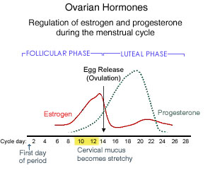
(FSH) and lutenizing hormone (LH). FSH
tells the granulosa cells to make LH receptors on the cell surface so that when
LH is produced and binds to the receptors, the end of the cycle proliferation
occurs. This makes the period stop (see
Figure 3).
Another
human gonadotropin is produced by the placenta, and this is known as human
chorionic gonadotropin (hCG). The hCG is
the hormone test for pregnancy that is commonly used on pregnancy strips. If hCG is present, placenta is making
it. As the placenta increases in size
during the early stages of pregnancy, the hCG also increases in number. During pregnancy, the placenta also produces
estrogen.
During
the nonpregnancy state, the human female ovaries produce estrogen,
progesterone, and testosterone.
Granulosa cells in the ovarian follicles and the surrounding corpora
lutea make estrogen. Other organ cells
participate in estrogen production, but to a lessor extent: the fat or adipose, liver, breasts, and the
adrenal gland. Postmenopausal estrogen
production can still occurs from these extra-ovarian sources, but a woman's individual blood levels must be measured to know what phase her ovaries are in. In the nonpregnant female, the highest levels
of estrogen occur just prior to ovulation, near the end of the Follicular Phase
(see Figure 3).

Figure 4. The metabolism of cholesterol. A variety of biochemical reactions exist whereby cholesterol is metabolized to progesterone, then on to dehydroepiandrosterone, testosterone, dihydrotestosterone, or estradiol. Cholesterol is not all bad, and our bodies must produce cholesterol not just in order to procreate, but to develop neurologically. Cholesterol is important to the structure of cells, as well as being a precursor of oxysterols, bile acids, and steroid hormones.
Cholesterol is the "Mother Molecule" of androgen and estrogen steroids (See Figure 4). Actually,
you may be surprised to learn that the cholesterol molecule is a major part of
the human brain, and there is no organ in the human body that contains more
cholesterol than the human brain (Orth, 2012).
In fact, about 20% of the body’s cholesterol is contained in the
brain. The brain does not have the same
metabolic pathway as other organs, and the brain is responsible for what is
called de novo synthesis of
cholesterol. This means that the brain
makes it freshly. It was Couerbe who, in 1836, described the cholesterol
molecule as being “un element principal”, meaning ‘a key element’ in the
central nervous system (Couerbe, 1834).
It is important to note that the ovary is uniquely tied into the hormones that they produce. The ovaries are an organ, and they synthesize and coordinate the lifecycle of a girl and a woman. In old age, the same ovaries dictate how menopause is approached.
If a woman undergoes a hysterectomy and the surgeon also removes the ovaries, this is 'surgical menopause'. A woman undergoing a hysterectomy gets a 'crash course' in menopause if the ovaries are removed, and she should be offered a discussion of whether or not she should be placed on hormone replacement therapy (HRT). Backing up for a moment, wait just one moment. Actually, we must first question whether the ovaries should be removed at all. Stay tuned for the next article, which will address this issue.
If a woman undergoes a hysterectomy and the surgeon also removes the ovaries, this is 'surgical menopause'. A woman undergoing a hysterectomy gets a 'crash course' in menopause if the ovaries are removed, and she should be offered a discussion of whether or not she should be placed on hormone replacement therapy (HRT). Backing up for a moment, wait just one moment. Actually, we must first question whether the ovaries should be removed at all. Stay tuned for the next article, which will address this issue.
References:
Couerbe
JP. Du cerveau, considere sous le point du vue chimique et physiologique.
Annales De Chimie Ed De Physique. 1834;56:160-193.
Orth
M., and Bellosta S. Cholesterol: its
regulation and role in central nervous system disorders. Cholesterol, 2012;2012:292598, doi:10.1155/2012/292598.
Epub 2012 Oct 17. http://www.ncbi.nlm.nih.gov/pubmed/23119149
~~~~~~~~~~~~~~~~~~~~~~~~~~~~~~~~~~~~~~~~~~~~~~~~~~~
To Order Dr. Margaret Aranda's Books, please go to:
http://drmargaretaranda.tateauthor.com/other-works/
http://drmargaretaranda.tateauthor.com/other-works/
To Order Dr. Aranda's Books and/or Pre-Order Archives of the Vagina
please click here: http://drmargaretaranda.tateauthor.com/other-works/
Face Book Page: No More Tears: A Physician Turned Patient Inspires Recovery
No More Tears en Espanol
Face Book Page: Stepping from the Edge
Little Missy Two-Shoes Likes to go to School
Face Book Page: Little Missy Two-Shoes Likes a Ladybug
From Menarche to Menopause: A Journey through Time
~~~~~~~~~~~~~~~~~~~~~~~~~~~~~~~~~~~~~~~~~~~~~~~~~~~~~~~~~~~~~~~~~~~~~~~~~~~~
Additional Free Articles by Dr. Margaret Aranda
Medical Disclaimer: Nothing on this website is meant to diagnose, treat, or practice medicine. You must be seen in person by a physician for appropriate and individual medical treatment. If you have an emergency, call 9-1-1 in the USA.
Link Disclaimer: We are not responsible for any links that go outside of this website.
Full Disclosure: Margaret A. Ferrante, M.D. is an Institute Physician with Cenegenics Medical Institute. She receives no monetary compensation for hosting this website you are on, which is independent and not affiliated with Cenegenics. The information presented is for education and awareness. Dr. Ferrante currently sees patients out of the Cenegenics office in Beverly Hills, CA.
To book an appointment for a free Consultation, please email her at: mferrante@cenegenics.com


Thank you for saying that women's reproductive organs are needed for more than just reproduction. When women do not understand this, they are much more likely to agree to hysterectomy. Women need to know that their reproductive organs are SEX organs and that there will never be a time they won't need them.
ReplyDeleteIn the most drastic comparison, one can not help but wonder how the masses of men would take it if their doctors told them, when referring to the testicles, "Well, you don't need them anyway." The ovaries and the testicles are representations of femininity and masculinity, respectively, and have a great deal to do with how each sex views themselves sexually.
ReplyDeleteDr. Margaret Aranda
www.drmargaretaranda@yahoo.com